Our Services

Radiology Associates offers a variety of services.

Choose a service to view information, frequently asked questions and suggested precautions prior to your examination.


FAQ

IDENTIFICATION

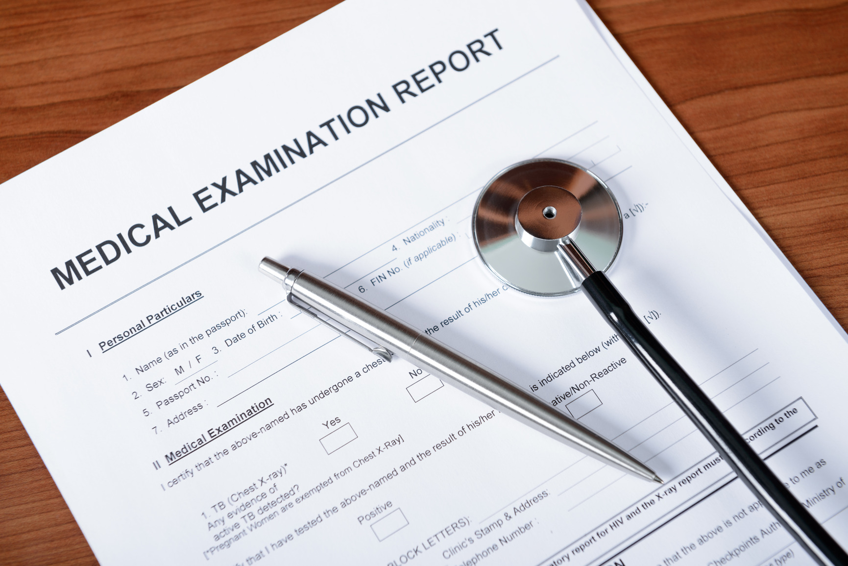
When you attend our office for an appointment, you need to bring the requisition you received from your doctor and present it to the front desk. At the request of Alberta Health Care, you are required to present your Alberta Health Care Card along with a piece of photo identification, such as a driver’s license or passport.

CANCELLATIONS

Please ensure that you cancel or reschedule at least 2 hours before your appointment start time. Having at least 2 hours notice that you will not be attending allows our office to contact another patient to attend in your place. This helps us to minimize wait lists.

CHILD CARE

We recognize that from time to time patients may be unable to arrange childcare when you come for your appointments. For those exceptional situations, we are pleased to report that you can now book your ultrasound in a specific room where children and other family members are allowed. Please remember that these services must be confirmed at the time of booking, and as there are limited numbers of these spots available, appointment times may be less flexible. Call us for more information.

WHEN TO ARRIVE

Please arrive at least 15 minutes before your scheduled appointment to allow us to obtain all pertinent information for your examination.

Typical Length of Exam
Approximately 20-30 minutes
Dress Requirements
Wear loose, comfortable clothing with no zippers, metal buttons, or other metal parts. Do not wear jewelry. Refrain from wearing perfume or cologne. Examination gowns are provided as necessary.
Medication and Dietary Requirements
You may take your usual medications except for calcium-containing medications on the day of the exam. If you have had a barium examination, intravenous contrast for a CT examination, or an isotope injection for a nuclear medicine examination, you will have to wait 10-14 days before having a body composition analysis.
SPECIAL ALERT
Be sure to tell your doctor or the technologist before the procedure if you are pregnant or believe you could be pregnant. This test is not usually performed during pregnancy.
Doctor referral required

Body Composition

Your body is made up of water, fat, protein, carbohydrates, vitamins, and minerals. Body composition analysis is used to determine the ratio of fat to lean body tissue. If you have too much fat, especially at the waist, you have a higher risk of health problems. Our facility uses dual energy X-ray absorptiometry (DEXA), a low dose X-ray procedure that is the best method for measuring body composition.
The information from this test can assist: 1. Physicians treating patients with obesity, cardiovascular disease, and other related health problems 2. Athletes and fitness enthusiasts who wish to monitor effects of training and exercise
You will be asked to lie on the examination table. A detector will move over your body and collect the information for a detailed body composition analysis. If you have any concerns or questions about the results, please discuss it with your physician or fitness instructor.
Typical Length of Exam
Approximately 20-30 minutes
Dress Requirements
Wear loose, comfortable clothing with no zippers, metal buttons, or other metal parts. Please do not wear jewelry. Refrain from wearing perfume or cologne. Examination gowns are provided as necessary.
Medication and Dietary Requirements
You may take your usual medications except for calcium-containing medications on the day of the exam. If you have had a barium examination, intravenous contrast for a CT examination, or an isotope injection for a nuclear medicine examination, you will have to wait 10-14 days before having bone mineral densitometry performed.
SPECIAL ALERT
Be sure to tell your doctor or the technologist before the procedure if you are pregnant or believe you could be pregnant. This test is not usually performed during pregnancy.
Doctor referral required

Bone Densitometry

Bone mineral densitometry, also known as dual energy X-ray absorptiometry (DEXA), is a low-dose X-ray examination used to assess for possible bone loss (osteoporosis).
  • Diagnosing osteoporosis
  • Determining fracture risk
  • Measuring the effects of treatment (It is best to perform this examination at the same facility every time to allow for more accurate comparison from one exam to the next.)
The technologist will ask you to lie on the examination table. For each body part to be examined, you will be positioned appropriately. Usually the lumbar spine and hips are examined but, occasionally, the forearm or total body may be assessed. A detector will move over the body part and an image will be generated on the computer monitor. A radiologist will then review the images and send a report to the referring physician.
Typical Length of Exam
This exam typically takes 60 minutes
Dress Requirements
Wear loose, comfortable clothing. Please do not wear jewelry. Refrain from wearing perfume or cologne. Examination gowns are provided as necessary.
Medication and Dietary Requirements
No special preparations are necessary.

Echocardiography

Echocardiography, often referred to as cardiac echo or simply echo, uses ultrasound to image the heart. Ultrasound is a safe and painless way to see the organs and tissue inside the human body by using very high frequency sound waves. The sound waves enter the body and bounce back at different rates depending on how they are absorbed by the body. When the sound waves bounce back, they are captured by the ultrasound machine and turned into a picture.  Echocardiography uses two-dimensional, three-dimensional and doppler ultrasound to create images of the heart.
Echocardiography is used to evaluate the heart muscle and its ability to pump, the valves of the heart, and the tissues surrounding the heart.
The ultrasound technologist will spread gel on your chest and then press a device known as a transducer against your skin, aiming the ultrasound waves toward your heart. This is a painless procedure; you will only feel the pressure of the transducer against your skin. As the scan continues, you may be asked to hold your breath or change position to give the best image possible. The technologist will then show the images to the radiologist to review all the images. Once the review is complete, the radiologist will send a report to, and may call, the doctor who ordered your test.
Typical Length of Exam
Approximately 45 minutes. Breast implants require more images and will require additional time. Please notify Radiology Associates when booking if you have implants.
Dress Requirements
On the day of the exam, please wash off all deodorants, perfumes, powders and/or lotions under the arms and across the chest. Wear loose comfortable clothing that can be removed from the waist up. Examination gowns are provided.
Medication and Dietary Requirements
No special preparations are necessary.
SPECIAL ALERT
Be sure to tell your doctor or the technologist before the procedure if you are pregnant or believe you could be pregnant. This test is not usually performed during pregnancy.

Breast Imaging

A mammogram is an X-ray picture of the soft tissues inside the breast. Mammograms require very small doses of radiation. The risk of harm from this radiation exposure is extremely low. The benefits of mammography almost always outweigh the potential harm from the very small amount of radiation.
There are two type of mammograms:
  • Screening Mammogram Mammograms can be used to check for breast cancer in women who have no signs or symptoms of the disease. This type of mammogram is called a screening mammogram. Screening mammograms usually involve two X-ray pictures, or images, of each breast. The X-ray images make it possible to detect tumours that cannot be felt. Screening mammograms can also find microcalcifications (tiny deposits of calcium) that sometimes indicate the presence of breast cancer.
  • Diagnostic Mammogram Mammograms can also be used to check for breast cancer after a lump or other sign or symptom of the disease has been found. This type of mammogram is called a diagnostic mammogram. Besides a lump, signs of breast cancer can include breast pain, thickening of the skin of the breast, nipple discharge or a change in breast shape or size. These changes can also be caused by benign conditions. A diagnostic mammogram can also be used to evaluate changes found during a screening mammogram, or to view breast tissue when it is difficult to obtain a screening mammogram because of special circumstances, such as the presence of breast implants.
You will be asked to remove your clothes from the waist up and will be given a gown to wear. You may find it easier to wear a blouse or shirt with pants or a skirt. X-rays will be taken of your breasts. To get a clear picture, your breast will be squeezed. This can cause some discomfort but is not harmful. Usually, two X-rays are taken of each breast, one from the side and one from the top. After you have completed your mammogram at Radiology Associates, a radiologist will review your images and compare them to any previous images that are available. The radiologist will then issue a report to your referring doctor. Sometimes the radiologist will ask for additional images for clarification or occasionally will ask for an ultrasound. This may happen on the same day as your initial mammogram or you may be asked to come back on another day for these additional images.
Screening mammography can help reduce the number of deaths from breast cancer among women ages 40 to 74 or in younger women who have a strong family history of breast cancer. Screening recommendations are as follows:
  • For women
  • For women 40-49 years old: screening mammogram every year.
  • For women 50-74 years old: screening mammogram every 2 years.
  • For women 75 years or older: screening mammogram every 2-3 years if desired.
Tomosynthesis is type of mammogram that produces 3-dimensional images of the breast. This allows for better detection of cancer as well as fewer call-back studies.
Whole breast ultrasound (also referred to as Automated Breast Ultrasound or ABUS) is used in addition to mammography to help in detecting breast cancers in women with dense breast tissue, or those who are at increased risk for breast cancer.
Typical Length of Exam
Approximately 45 minutes to 1 hour
Dress Requirements
Wear loose clothing without metal or buttons (e.g. sweatpants, track pants, or yoga pants.) Please do not wear jewelry. Refrain from wearing perfume or cologne. Examination gowns provided as necessary.
Medication and Dietary Requirements
No dietary restrictions. As tolerated, please reduce intake of oral pain killers on the day of your appointment. You will need to report any allergies, chance of pregnancy, medication such as blood thinners or antibiotics, and medical conditions such as diabetes.
SPECIAL ALERT
Be sure to tell your doctor or the technologist before the procedure if you are pregnant or believe you could be pregnant. Pain procedures that use X-rays are not usually performed during pregnancy. Pain procedures using ultrasound can still be performed.

Pain Clinic

These minimally invasive procedures are performed by a radiologist using X-ray or ultrasound guidance to precisely deliver anaesthetic and anti-inflammatory medications to specific areas causing pain, including joints, bursae, tendons, nerve roots, etc. The goal is to improve the patient’s quality of life by decreasing pain and increasing functional ability.
A wide variety of patients may benefit from these services including those suffering from pain due to arthritis, previous trauma, sports injuries, and sciatica, as well as patients awaiting joint replacement.
You will need a referral from your family physician or a specialist. Once you have a referral, call for an appointment. You will be asked several preliminary questions about your medical history and medications to ensure you can have the procedure safely.
The appointment may include a brief consultation to confirm the source of pain. Consent forms will explain the minimal risks and rare complications. Written consent will be required. It may be necessary to change into an examination gown. You will be positioned in either a chair or bed to allow the radiologist to visualize the target area with X-ray or ultrasound. The skin overlying the affected area will be sterilized and protected with surgical drapes. The radiologist will anesthetize the injection site, then the needle will be placed accurately using image guidance. You will be required to stay for a short time following the injection.
Immediate effects from the local anesthetic will usually last for several hours. Your initial response will be recorded with a pain score. Symptoms may then return for a short period of time until the anti-inflammatory medication has had sufficient time to work, usually within several days to a week. The effects will typically last for several months.
The Pain Management Clinic is located on the downstairs level of the Radiology Associates clinic in Lethbridge.
The majority of the procedures are free of charge to patients with valid Alberta Health Care insurance. Bring your Alberta Health Care Card and a piece of photo identification with you to the appointment.
Typical Length of Exam
Approximately 45 minutes to 1 hour
Dress Requirements
Wear loose clothing without metal or buttons (e.g. sweatpants, track pants, or yoga pants.) Please do not wear jewelry. Refrain from wearing perfume or cologne. Examination gowns provided as necessary.
Medication and Dietary Requirements
As tolerated, please reduce intake of oral pain killers on the day of your appointment. You will need to report any allergies, chance of pregnancy, medication such as blood thinners or antibiotics, and medical conditions such as diabetes.
SPECIAL ALERT
Be sure to tell your doctor or the technologist before the procedure if you are pregnant or believe you could be pregnant. Pain procedures that use X-rays are not usually performed during pregnancy. Pain procedures using ultrasound can still be performed.

Ultrasound

Ultrasound scanning is also known as sonography. Ultrasound exams are a safe and painless way to see the organs and tissue inside the human body by using very high frequency sound waves. The sound waves enter the body and bounce back at different rates depending on how they are absorbed by the body. When the sound waves bounce back, they are captured by the ultrasound machine and turned into a picture.
There are many uses for ultrasound. Some common ones are to assess the liver, spleen, gallbladder, pancreas, kidneys or bladder. Ultrasound is also a very good and very safe way to look at babies that are developing inside the uterus. It is routinely used to assess the breasts, thyroid, scrotum, neck, shoulder, elbow, wrist, knee, and ankle, and is used to look at arteries and veins throughout the body. Ultrasound is also used to look at the heart. Check out the Echocradiography section for more info on Ultrasound of the heart.
When you arrive for your test, you will be asked to remove all clothing and jewelry from the area being scanned. You will be given a hospital gown if needed and will be asked to sit or lie down on an examination table. A small amount of gel will be applied to the skin over the area to be scanned to help the sound waves move into your body. The ultrasound technologist will slide the small ultrasound instrument back and forth through this gel. The ultrasound instrument, also called a transducer, will transmit ultrasound waves into your body and receive their reflected echoes. This is a painless procedure; you will only feel the transducer against your skin. As the scan continues, you may be asked to hold your breath or change position to give the best image possible. The technologist will then show the images to the radiologist to review. Once the review is complete, the radiologist will send a report to, and may call, the doctor who ordered your test.
Unfortunately we cannot provide child care. Please make other arrangements for your child or have another adult present who can wait with your child in the waiting room. Fathers without children present are welcome to view an obstetrical ultrasound while it is performed. Fathers with children present will be asked to remain in the waiting room until the end of the exam before they will be brought in to view the baby. We recognize that from time to time patients may be unable to arrange childcare when you come for your appointments.  For those exceptional situations, we are pleased to report that you can now book your ultrasound in a specific room where children and other family members are allowed.  Please remember that these services must be confirmed at the time of booking, and as there are limited numbers of these spots available, appointment times may be less flexible.  Call us for more information.
Exam-Specific Instructions
  • Approximately 45 minutes
  • Do not eat or drink after midnight the night before your exam.
  • No chewing gum is permitted (gum generates saliva with small particles of air that, when swallowed, make organs harder to see).
  • Brushing teeth is permissible.
  • For infants, withhold last feeding prior to appointment time.
  • Medication can be taken at its usual time with a small amount of water.
  • Only patients will be allowed in the exam room unless caregivers are needed.
  • Approximately 60 minutes
  • Do not eat or drink after midnight the night before your exam. 90 minutes before your appointment, empty your bladder and then finish drinking four 8 oz. glasses of water (1 litre total).
  • DO NOT VOID within 90 minutes of your appointment.
  • DO NOT SUBSTITUTE WITH ANY OTHER LIQUID.
  • Children (12 and under) are only required to drink 2 glasses of water, 8 oz. each (500 mL total).
  • Only patients will be allowed in the exam room unless caregivers are needed.
  • Approximately 30 minutes
  • 90 minutes before your appointment, empty your bladder, then finish drinking four 8 oz. glasses of water (1 litre total).
  • DO NOT VOID within 90 minutes of your appointment.
  • DO NOT SUBSTITUTE WITH ANY OTHER LIQUID.
  • Children (12 and under) are only required to drink 2 glasses of water, 8 oz. each (500 mL total).
  • Only patients will be allowed in the exam room unless caregivers are needed.
  • Approximately 45 minutes
  • 90 minutes prior to your appointment, empty your bladder, then drink water as specified below. The amount of water you need to drink depends on how far along you are in your pregnancy: Up to 25 weeks – 3 glasses of water, 8 oz. each (750 mL total) Over 25 weeks – 1 glass of water, 8 oz. (250 mL total)
  • DO NOT VOID within 90 minutes of your appointment.
  • DO NOT SUBSTITUTE WITH ANY OTHER LIQUID.
  • Fathers without children present are welcome to view the obstetrical ultrasound while it is performed. Fathers with children present will be asked to remain in the waiting room until the end of the exam when they will be brought in to view the baby.
  • USB sticks with ultrasound images of your baby are available for purchase following your exam.
  • Upper extremities (No preparation)
  • Lower extremities (Nothing to eat or drink after midnight)
  • Renal arteries (Nothing to eat after midnight, drink two glasses of water two hours before your appointment, a full bladder is not required)
No special preparation required.
Typical Length of Exam
An initial vein mapping ultrasound will take around 60 minutes. A second appointment will be made for consultation to discuss the ultrasound results and your treatment options. This will also take approximately 60 minutes.
Dress Requirements
Wear loose clothing without metal or buttons (e.g. sweatpants, track pants, or yoga pants.) Please do not wear jewelry. Refrain from wearing perfume or cologne. Examination gowns provided as necessary.
Medication and Dietary Requirements
For the vein mapping ultrasound appointment, no preparation is required. For the consultation appointment, please bring your list of current medications and allergies. If undergoing a treatment, do not use any products containing aspirin 4 days prior to the procedure, and eat a small meal 1 to 2 hours prior to arriving at the clinic.
SPECIAL ALERT
If undergoing a vein procedure, please arrange for transportation following your appointment as you will be receiving a local anesthetic/oral sedative and WILL NOT BE ABLE TO DRIVE. Also, be advised that long trips are not permitted 4 to 7 days following your surgery. If trips are unavoidable, get out and walk hourly.

Vein Clinic

Chinook Vascular is located at the Radiology Associates clinic in Lethbridge. Please come to the main reception desk when you arrive.
Chinook Vascular is a subgroup of Radiology Associates which focuses on the diagnosis and treatment of venous insufficiency. Diagnosis is made through patient history, physical exam and ultrasound evaluation. A variety of therapeutic options are offered such as compression stockings, injections, endovenous laser ablation, and referral for surgery. Procedures are performed onsite in an accredited non-hospital surgical suite.
Venous insufficiency occurs when your veins have trouble sending blood from your limbs back to your heart. Symptoms of venous insufficiency may include varicose veins, spider veins, burning or itching in the legs, and ankle swelling and ulceration.
An ultrasound map of the venous system may be ordered by your physician. Subsequently, an appointment may be made to see a nurse for simple cosmetic injections. For more serious complaints, a consultation may be ordered by your physician. At that time you will watch a video, meet with one of our radiologists, have a history taken and undergo a physical exam. Therapeutic options and their risks will be discussed.

It is estimated that 25% of women and 15% of men suffer from venous insufficiency. This can result in:

  • Painful legs
  • Bulging veins
  • Swelling
  • Aching
  • Spider veins
  • Tingling
  • Burning
  • Throbbing
  • Cramping
  • Itchiness
  • Leg fatigue
  • Leg heaviness
  • Restless legs
  • Discoloration
  • Inflammation
  • Ulceration

Chinook Vascular currently offers the following therapeutic options for venous insufficiency:

  • Endovenous laser ablation
  • Foam echosclerotherapy
  • Liquid sclerotherapy
  • Compression stockings

Endovenous laser ablation is a “state of the art” minimally invasive alternative to conventional surgical stripping of veins. If this procedure is determined to be an appropriate treatment for you, you can rest assured knowing that it is quick and effective, and can treat both the pain and appearance of varicose veins. Most patients are able to return to normal activities within a week.

Chinook Vascular is an accredited, non-hospital surgical facility.

Contact your family physician for a referral.

If undergoing a vein procedure, please arrange for transportation following your appointment as you will be receiving a local anaesthetic/oral sedative and WILL NOT BE ABLE TO DRIVE. Also, be advised that long trips are not permitted 4 to 7 days following your surgery. If trips are unavoidable, get out and walk hourly.
Typical Length of Exam
Approximately 5-25 minutes depending on the exam type.
Dress Requirements
Wear loose, comfortable clothing. You may need to remove your glasses, watch, rings or other metal that may be in the way of the part being X-rayed. Please refrain from wearing perfume or cologne. Examination gowns are provided as necessary.
Medication and Dietary Requirements
No special preparations are necessary.
SPECIAL ALERT
Be sure to tell your doctor or the technologist before the procedure if you are pregnant or believe you could be pregnant. This test is not usually performed during pregnancy.

X-Ray

X-ray is a common imaging test that has been used for decades to help doctors view the inside of the body. X-rays use small amounts of radiation. The level of exposure is considered safe for adults; however, it is not generally considered safe for a developing fetus. Be sure to tell your doctor or the X-ray technologist before the procedure if you are pregnant or believe you could be pregnant.
X-ray imaging is useful for looking at bones for fractures or joint problems including arthritis, as well as for assessing some tumours or infections. It is used to look at problems affecting the neck, lungs or heart. X-rays are also good for looking at digestive problems or items that have been swallowed.
The technologist will most likely require you to lie, sit, or stand in several positions during the test. Some images may be taken while you stand in front of a specialized plate that contains X-ray sensors. You will have to hold your breath and remain still while the images are being taken. This provides the clearest images possible. The technologist will position your body in such a way as to get the best picture of the part your doctor wants to see. You can help by following the instructions you are given. The technologist will then go into the small room with the controls, located adjacent to the X-ray room. They will push a button to take the picture and you will probably hear a clicking noise and a beep. This process is repeated until the technologist has acquired all the images needed. A radiologist will review the images and send a report to the referring physician.